


Cells 2024, 13(9), 729; https://doi.org/10.3390/cells13090729 (registering DOI) - 23 Apr 2024
Abstract
Fibrous dysplasia (FD) is a rare bone disorder characterized by the replacement of normal bone with benign fibro-osseous tissue. Developments in our understanding of the pathophysiology and treatment options are impeded by the lack of suitable research models. In this study, we developed
[...] Read more.
Fibrous dysplasia (FD) is a rare bone disorder characterized by the replacement of normal bone with benign fibro-osseous tissue. Developments in our understanding of the pathophysiology and treatment options are impeded by the lack of suitable research models. In this study, we developed an in vitro organotypic model capable of recapitulating key intrinsic and phenotypic properties of FD. Initially, transcriptomic profiling of individual cells isolated from patient lesional tissues unveiled intralesional molecular and cellular heterogeneity. Leveraging these insights, we established patient-derived organoids (PDOs) using primary cells obtained from patient FD lesions. Evaluation of PDOs demonstrated preservation of fibrosis-associated constituent cell types and transcriptional signatures observed in FD lesions. Additionally, PDOs retained distinct constellations of genomic and metabolic alterations characteristic of FD. Histological evaluation further corroborated the fidelity of PDOs in recapitulating important phenotypic features of FD that underscore their pathophysiological relevance. Our findings represent meaningful progress in the field, as they open up the possibility for in vitro modeling of rare bone lesions in a three-dimensional context and may signify the first step towards creating a personalized platform for research and therapeutic studies.
Full article
(This article belongs to the Special Issue Model Systems for Human Disease and Medicine: From Advanced Cell and Tissue Culture to In Silico Methods)
►
Show Figures
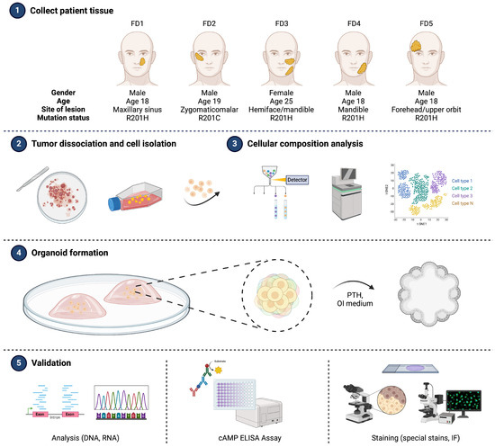
Figure 1


